Continuing Education
COURSE FINDER
Please fill out at least one form field to perform a search.

Biological Imaging: Microscopes in Art
Mar 22 - Apr 26
$350
Location
335 West 16th Street
Faculty
Joseph DeGiorgis,
Marine biologist
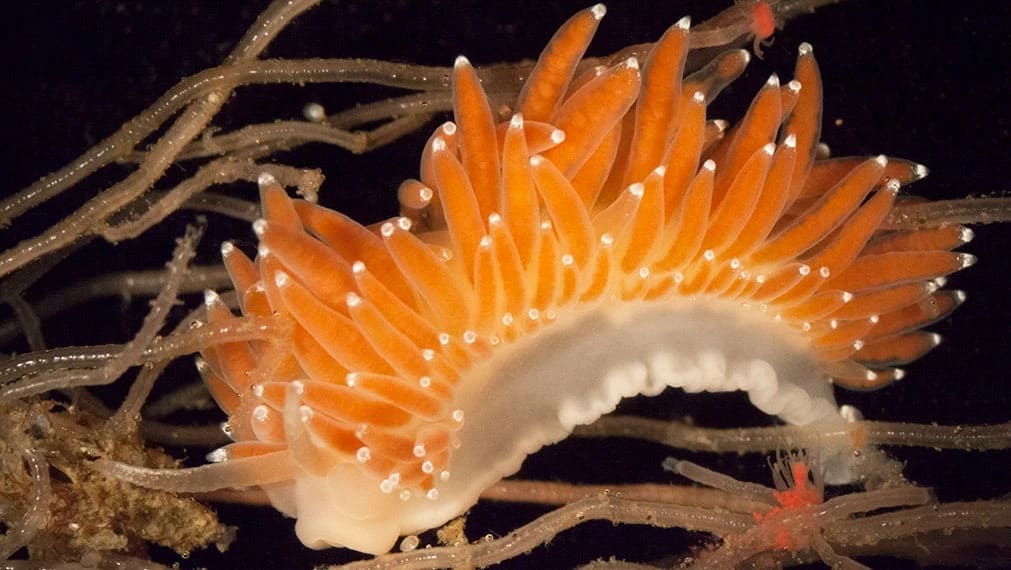
The light microscope was first developed and famously used in the late 1600s by the Dutch naturalist, Antonie van Leeuwenhoek, to look at small pond creatures he called "animalcules." Observations with these instruments also led to the discovery of cells as the basic unit of life, one of the foundations of biological science. Microscopes remarkably make the invisible visible through magnification, allowing for the observation and photo documentation of small forms of life and tiny features of larger organisms. These forms are often beautiful and exhibit morphologies beyond one's dreams and imagination. Capturing images of these anatomies transcends science into the realm of art and into our society and culture. In this course we will use a variety of microscopes to take still and moving images of bacteria, plankton, protozoa, lichens, fungi, marine invertebrates, flowers, and other botanical specimens. Each student will create an artist's portfolio of photographs and short movies of microscopic life. All equipment and materials will be provided.
NOTE: Students will need a DSLR memory card or an external hard drive to store their projects. This course is held on campus at SVA.
NOTE: Students will need a DSLR memory card or an external hard drive to store their projects. This course is held on campus at SVA.
Course Number
FIC-2516-B
Credits
2 CEUs


